Services » HomeServices
Radiology :-
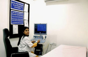
USG
Ultrasound technique of diagnosis uses sound waves very similar to the sonics used by the bats and sophisticated jets to locate an object.
Different human body organs respond to these waves differently and this response facilitates the diagnosis of the normality and abnormality of the tissues.
It is most commonly used for foetal studies, abdominal, thyroid, breast, orbits, and testes studies etc.
It is the safest technique of diagnostics using harmless waves as a tool for detection
Time taken: 30 – 90 min depending upon the area
3D ULTRASOUND
Using ultrasound 3D images can be obtained of different parts of the body to assess their well being. It is especially beneficial in detecting foetal anomalies by using this scan.
The joy of new life is unmatched and immense and the assured safety of this little being is the biggest concern for everybody.
We provide a safe harbour in the form of our facility.
In 3D fetal scanning, however, instead of the sound waves being sent straight down and reflected back, they are sent at different angles. The returning echoes are processed by a sophisticated computer program resulting in a reconstructed three-dimensional volume image of the fetus’s surface or internal organs, in much the same way as a CT scan machine constructs a CT scan image from multiple x-rays.
Time taken: 30 – 90 min
Digital x ray
The x rays taken using digital receptors and computers instead of normal x ray films are called digital x rays.
The common digital method used is the CR system that produces digital X-rays on a computer.
They have better quality and contrast and thereby facilitate better and accurate diagnosis.
Colour Doppler
Colour Doppler is a specialised Ultrasonography technique using sound waves to evaluate the flow and velocity of the blood in veins and arteries.
Using the Doppler principle of changing pitch with velocity, ultrasound waves that reflect from the red blood cells in the arteries and veins are evaluated for velocity using which amplitude and coloured maps of the vessels are created.
It is very helpful in the study of carotid artery of the neck, the abdominal arteries and that of upper and lower limbs.
Time taken – 30 – 60 min depending upon the site.
2D Echo
Heart disease is a number one cause of death worldwide causing 17.3 million deaths and over 80% deaths occur in developing countries like ours. Timely diagnosis and treatment prevents the burden of the disease to a large extent and can save a lot many lives.
Doppler echocardiography is a procedure that uses ultrasound technology to examine the heart or blood vessels. An echocardiogram uses high frequency sound waves to create an image of the heart and allows determination of the speed and direction of blood flow by utilizing the Doppler Effect.
An advantage of Doppler echocardiography is that it can be used to measure blood flow within the heart without invasive procedures such as cardiac catheterization.
We offer this facility dedicated to our fellow society members……..
Time taken: 45 mins
DXA scan
Bone density scanning, also called Dual-Energy X-ray Absorptiometry (DXA) or bone densitometry, is an enhanced form of x-ray technology that is used to measure bone loss. DXA is today’s established standard for measuring bone mineral density (BMD).
DXA is most often performed on the lower spine and hips. In children and some adults, the whole body is sometimes scanned.
A screening is done through the BMD screening device and further advance scans can be done using DXA.
A scan is recommended for you if –
You are a post-menopausal woman and not taking estrogen.
Have a personal or maternal history of hip fracture or smoking or with clinical conditions associated with bone loss.
Use medications that are known to cause bone loss, including corticosteroids such as Prednisone, various anti-seizure medications such as Dilantin and certain barbiturates, or high-dose thyroid replacement drugs.
Have type 1 (formerly called juvenile or insulin-dependent) diabetes, liver disease, kidney disease or a family history of osteoporosis.
Have a thyroid condition, such as hyperthyroidism.
Have a parathyroid condition, such as hyperparathyroidism.
Have experienced a fracture after only mild trauma.
Have had x-ray evidence of vertebral fracture or other signs of osteoporosis.
Time taken – 30 min
Mammography
We offer Digital mammography, also called full-field digital mammography (FFDM). Digital mammography is a specialized form of mammography that uses digital receptors and computers instead of x-ray film to help examine breast tissue for breast cancer.
It improves the image quality and contrast and hence facilitates better diagnosis as accuracy in diagnosis of breast diseases is extremely important for timely treatment options planning.
Mammography is done as –
Screening mammography:
Screening Mammography plays an extremely important role in the early detection of breast cancers because it can show changes in the breast up to two years before a patient or physician can feel them. The American Cancer Society (ACS), the American Medical Association (AMA) and the American College of Radiology (ACR) recommend screening mammography every year for women, beginning at age 40. So they can be treated while they most curable.
Diagnostic Mammography
Diagnostic mammography is used to evaluate a patient with abnormal clinical findings—such as a breast lump or lumps—that have been found by the woman or her doctor.
Diagnostic mammography may also be done after an abnormal screening mammogram in order to evaluate the area of concern on the screening examination.
Time taken – about 30 – 40 min each side.
OPG
OPG stands for Orthopantomography. It is a special method for obtaining foolscap images of the both upper and lower jaws where in all the teeth as well as the bone surrounding them can be visualised.
A very important tool for dental diagnosis and treatment, all treatment options whether in oral surgery, orthodontics, periodontics, prosthetics or implants require this as a must.
Technology: a specialised x-ray emitting machine which rotates around the head to capture the full picture of the jaws and the teeth.
Time taken: About 20 min
Digital IVP
An intravenous pyelogram (IVP) is an x-ray examination of the kidneys, ureter and urinary bladder that uses iodinated contrast material injected into veins.
When a contrast material is injected into a vein in the patient’s arm, it travels through the blood stream and collects in the kidneys and urinary tract, turning these areas bright white on the x-ray images. An IVP allows the radiologist to view and assess the anatomy and function of the kidneys, ureters and the bladder.
Time taken – About 1.5 hr.
Barium scan
Barium tests are used to help see the outline of the upper parts of the gut (gastrointestinal tract) such as the oesophagus, stomach and small intestines. It is extensively utilised in identification of abnormalities in the lining of the gut, any tumours, ulcers etc.
The gut (gastrointestinal tract) does not show up very well on ordinary X-ray pictures. However, when a white liquid containing a chemical called barium sulphate is consumed,
the outline of the upper parts of the gut (oesophagus, stomach and small intestines) shows up clearly on X-ray pictures.
Types of barium tests – Barium swallow, barium meal, Barium follow through, small intestine barium enema, colon barium enema.
Time taken – 20 – 50 min depending upon the area to be examined.

Copyright © 2018 All Rights Reserved Mammography| Cancer screening| Blood testing| Diagnostic centre| Jaipur